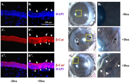
Fig. 9.
Expression of ΔE3β-catenin alone can induce corneal epithelial neoplasia in adult mice. (A-B″) Krt12rtTA/Wt/tetO-Cre/Ctnnb1floxE3/Wt mice induced without (−Dox) (A,A′,A″) or with Dox (+Dox) (B,B′,B″) for three days. Mouse eyes were enucleated and prepared for paraffin blocks. Corneal sections were subjected to immunofluorescence staining with anti-β-catenin antibody (red) and counterstained with DAPI (blue). Wild-type β-catenin was presented on the cell membrane of non-induced cornea (A,A′,A″), whereas Dox-induced expression of ΔE3β-catenin was found in nuclei (arrowheads in B′,B″) and was tightly associated with the mis-arrangement of the corneal epithelium to form corneal nodules (arrows in B,B′,B″). (C-H) As compared with un-induced mice that exhibited transparent cornea (C,D), four out of four Krt12rtTA/Wt/tetO-Cre/Ctnnb1floxE3/Wt mice induced with Dox (from P21-P35) developed OSSN (E-H) with leukoplakic pannus-like lesions (arrowheads in E,H) in the cornea with neovascularization (arrows in E,F). D, F and H represented higher magnification images from C, E and G, respectively.

