Fig. 1.
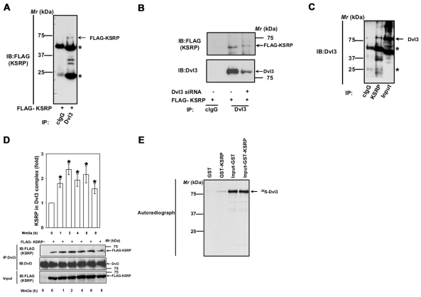
KSRP interacts with Dvl3. (A) F9 cells were transiently transfected with FLAG-KSRP for 24 hours followed by cell lysis and affinity pull-downs with either mouse control IgG or anti-Dvl3 mouse monoclonal antibody. Interaction of KSRP with Dvl3 was visualized by probing the blots with anti-FLAG antibody. Asterisks indicate the bands of immunoglobulin heavy and light chains. (B) F9 cells were treated with 100 nM of Dvl3 siRNA for 24 hours followed by transient expression of FLAG-KSRP for 24 hours followed by cell lysis and affinity pull-downs with either mouse control IgG or anti-Dvl3 mouse monoclonal antibody. Interaction of KSRP with Dvl3 was visualized by probing the blots with anti-FLAG antibody. (C) F9 cell lysates were immunoprecipitated with either rabbit control IgG or rabbit anti-KSRP polyclonal antibody and the interaction of KSRP with Dvl3 was visualized by probing the blots with anti-Dvl3 mouse monoclonal antibody. (D) F9 cells were transiently transfected with empty vector or FLAG-KSRP for 24 hours. The cells were then treated with Wnt3a (10 ng/ml) for indicated period of time followed by cell lysis and affinity pull-downs with anti-Dvl3 specific antibodies followed by immunoblotting with anti-FLAG antibodies. (E) To test the direct interaction of Dvl3 with KSRP, in vitro synthesized 35S-labeled Dvl3 was used in pull-down experiments with either GST- or GST-KSRP-Sepharose beads in the presence of 0.8% BSA. The interaction was visualized by SDS-PAGE and autoradiography. Representative blots of three independent experiments that proved highly reproducible are shown. *P<0.05 versus control (−Wnt3a).

