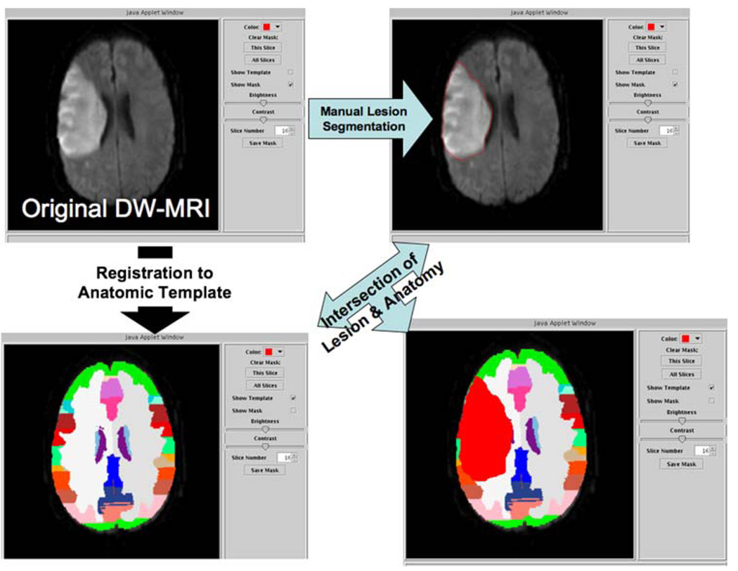
Fig. 2.
WebParc visualization interface. The user can visualize the clinical images (upper left), visually validate the registration between clinical case and template (lower left), and then proceed to make the lesion segmentation using mouse-driven cursor (upper right). Upon completion of the segmentation, the user initiates the intersection of the lesion mask with the template-based regions of anatomic interest, yielding a “lesion volume” for each anatomic region that the lesion intersects with (lower right)

