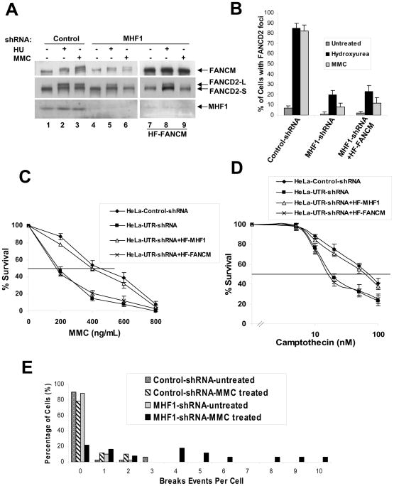
Figure 2. MHF1 is required for the activation of the FA pathway.
(A) Immunoblot showing that ablation of MHF1 expression in HeLa cells by shRNA reduces the level of monoubiquitinated FANCD2 in cells treated with MMC or HU. MHF1 knock down HeLa cells stably overexpressing HF-FANCM were used to show that phenotype observed is not due to the reduced levels of FANCM. (B) Histogram of immunofluorescence analysis of FANCD2 nuclear foci in HeLa cells in which MHF1 expression was knocked down by shRNA. Cells were either left untreated or exposed to MMC or HU, and the percentage of cells with 5 or more FANCD2 foci was determined by examining at least 150 cells. The result shows the average of 3 independent experiments with standard deviations. See also Fig. S2A. (C) & (D) Graph showing MMC and camptothecin survival curve of MHF1 knockdown cells. HeLa cells were transduced with either control or shRNA oligos targeting MHF1 and were subsequently treated with the indicated concentration of MMC and camptothecin. Visible colonies from 200 cells were counted after 10 days. The data represent the percent survival, as compared with untreated cells. Each experiment was performed in triplicate, and mean values are shown with standard deviations. HeLa cells stably expressing shRNA resistant HF-MHF1 were used as a control to show that the phenotype observed is not due to the off-target effect of shRNA. Also, MHF1 knock down HeLa cells stably overexpressing HF-FANCM were used to show that phenotype observed is not due to the reduced levels of FANCM. (E) Chromosomal aberrations following MMC treatment. HeLa cells were transfected with siRNAs and cells were treated with MMC. Metaphase spreads were prepared and scored for chromosomal aberrations. See also Fig. S2E.

