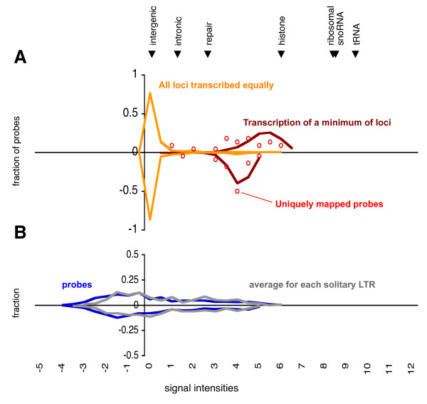
Figure 2.
HybMap signal intensities for LTR sequences. The distributions of signal intensities for HybMap probes mapping to full-length LTR retrotransposons (A) and solitary LTRs (B). General figure format as in Figure 1. A) Intensity distribution of full-length LTR probes are displayed using two different procedures: i) each probe's intensity divided by the number of possible mappings (assuming all LTR loci being transcribed equally; yellow curve), ii) total intensity of each probe assigned to one locus (transcription of a minimum of loci; red curve). The intensity distribution of probes mapping uniquely to a single full-length LTR retrotransposon locus are shown as open red circles. B) Intensity distribution for probes mapping uniquely to solitary LTR sequences (blue curve). For each solitary LTR loci the average intensity was calculated and plotted (grey curve). For comparison, the median intensity of forward probes mapping to other genomic features are indicated at the top of the figure.

