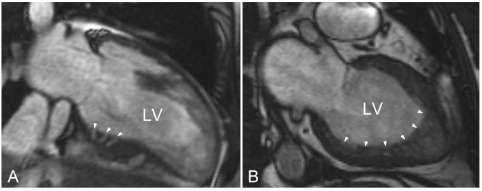
Figure 1.
Cardiac magnetic resonance cine images. A) Modified two-chamber end-diastolic cine image through the inferoseptum of a hypertrophic cardiomyopathy mutation carrier. Crypts are present in the basal inferoseptum (denoted by white arrowheads) penetrating compact myocardium and can easily be distinguished from noncompaction cardiomyopathy. B) Two-chamber cine imaging in a patient with noncompaction cardiomyopathy. The noncompacted layer aligning a compact layer is most profound in the apical region, and sometimes extends towards the inferior and/or lateral regions, as illustrated by the white arrowheads. LV= left ventricle.

