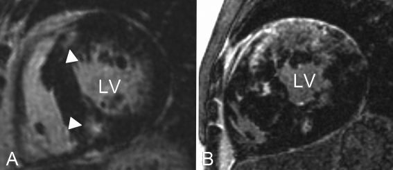
Figure 2 .
Short-axis left ventricular late gadolinium enhancement images of two HCM patients. Note that the fibrotic burden in patient B (confluent type LGE) is much more extensive than in patient A (patchy type LGE), which is related to increased risk of ventricular arrhythmias. Typically, late gadolinium enhancement (white arrowheads) is located at the insertion points of the right ventricle into the septum, see patient A. This pattern is observed in approximately 80% of HCM patients. LV=left ventricle.

