
Figure 2.
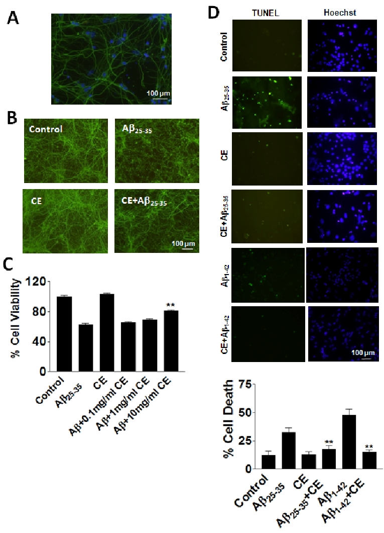
CE protects against Aβ toxicity in rat primary neurons. A. There are 90% neurons in rat primary prefrontal tissue cultures. Green: specific neuronal marker tubulin III; blue: Hoechst. B. Tubulin III staining shows the morphology of neurons under treatments. Aβ25-35 induces significant deform of neuronal morphology after 24 hours of treatment. CE (10 mg/ml) reverses the morphological changes induced by Aβ25-35. C. MTT assays show that CE (10 mg/ ml) increases cell viability compared with Aβ treatment. D. TUNEL assays show that CE (10 mg/ml) decreases cell death induced by Aβ. Data represent mean+SE (n=3). **P<0.01 compared to Aβ25-35 or Aβ1-42 group. Scale bar: 100 mm.