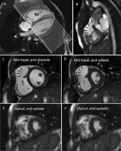
Figure 1.
Right ventricular volume measurement in CHD: several regions are challenging and need consistent approaches for comparison between studies. The images are from a patient after repair of tetralogy of Fallot with infundibular resection. (A) The short-axis stack (typically 6 mm slice thickness with 4 mm gaps) is shown relative to four-chamber cine. The most basal short-axis cine should be located just within the basal myocardium of the RV and LV at end diastole. However, the tricuspid annular plane (dotted line) may lie oblique to this slice, and usually moves through the first and often the second slice during systole. Care is needed to delineate areas of the ventricular but not the atrial cavities in the more basal slices. (B) A thin, akinetic region of the RVOT (arrowed in the sagittal RVOT view) should be regarded as part of the RV up to the (expected) level of the pulmonary valve. (C) In a mid-basal short-axis slice, the arrows indicate an akinetic region. A relatively smooth contour is drawn immediately inside the compact myocardium of the free wall, outside the trabeculations. (D) However, at end systole, hypertrophied trabeculations of the muscular part of the free wall may appear to merge (asterisk). The boundary line can still be located just inside the compact layer after viewing in cine mode. Alternatively, the dotted line drawn within the trabecular layer would give a slightly smaller end-systolic blood volume. (E) Trabeculations are numerous towards the apex of the RV and partial volume averaging blurs boundaries, so delineation outside the blood and trabeculations is probably the most reproducible approach. (F) Alternatively, tracing around the visible trabeculations at all levels may be more accurate, although not necessarily more reproducible between investigators and studies. The methods chosen need to be consistent for longitudinal comparison.

