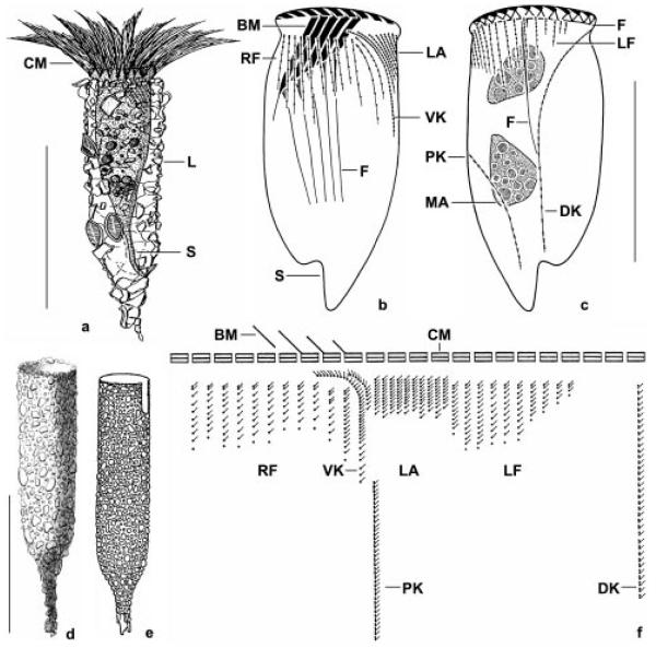
Figs 1a-f.
Tintinnopsis cylindrica (a-d, f) and a supposed synonym (e) from life (a, e), after protargol impregnation (b, c, f), and preserved with mercuric chloride (d). a - a representative specimen from the neotype population; b, c - ciliary pattern of ventral and dorsal side. Note the fibres that are associated with the oral and somatic ciliature; d - a lorica from the type population (from Daday 1887); e - Tintinnopsis kofoidi (from Hada 1932a); f - kinetal map of a morphostatic specimen. BM - buccal membranelle, CM - collar membranelles, DK - dorsal kinety, F - probably fibrillar structures, L - lorica, LA - lateral ciliary field, LF - left ciliary field, MA - macronuclear nodules, PK - posterior kinety, RF - right ciliary field, S - stalk, VK - ventral kinety. Scale bars: 100 μm (a, d, e); 50 μm (b, c).

