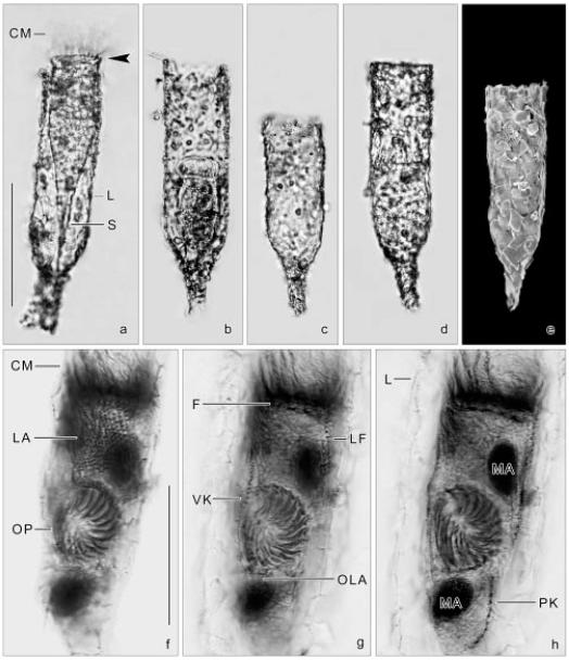
Figs 3a-h.
Tintinnopsis cylindrica from life (a-d), in the scanning electron microscope (e), and after protargol impregnation (f-h). a-d - lateral views showing lorica variability, concerning the degree of incrustrated particles and length of the cylindroidal portion. Accessory combs (arrowhead; a) are rarely recognizable; e - the lorica wall has many silt particles and fragments of diatom frustules incrustrated; f-h - same middle divider at three focal planes. The new oral apparatus develops in a subsurface pouch posterior to the proter’s lateral ciliary field. CM - collar membranelles, F - probably fibrillar structures, L - lorica, LA - proter’s lateral ciliary field, LF - left ciliary field, MA - macronuclear nodules, OLA - opisthe’s lateral ciliary field, OP - oral primordium, PK - posterior kinety, S - stalk, VK - ventral kinety. Scale bars: 100 μm (a-e); 50 μm (f-h).

