
Figure 7.
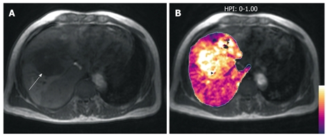
A middle-aged man with colorectal liver metastases to the liver. A: T1-weighted axial MR image demonstrates a hypointense liver metastasis in the right liver lobe (arrow); B: HPI map (calculated by the method described by Miles et al[2]) overlaid on the T1-weighted image shows increased HPI within the metastasis, typical of malignant disease.