
Figure 2.
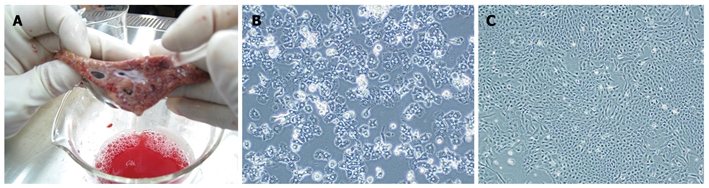
Isolation technique and microscopic appearance of primary and immortalized porcine hepatocytes. A: Perfusion was conducted by inserting a suitable pipette into vessels exposed on a cut surface of the sample; B: Non-immortalized primary porcine hepatocytes grow slowly with low plating efficiency and a short life span; C: Immortalized porcine hepatocytes grow rapidly as islands and had an extended life span. Magnifications: B and C 100 ×.