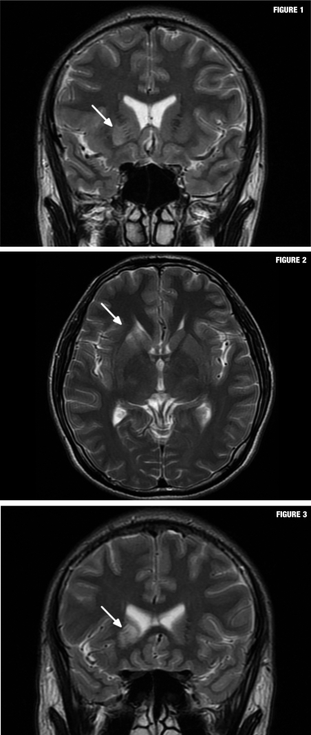
FIGURES 1–3.
Axial and coronal fast-spin echo T2-weighted images (1.5 Tesla, TR 4000ms, TE 125ms, slice thickness 5mm, 320 x 224 matrix, NEX 2) revealed a high signal intensity lesion in the lower portion of the head of the right caudate nucleus, inferior portion of the anterior limb of the right sided internal capsule, and the anterior third of the right putamen. The shape and location of this lesion matched the medial lenticulostriate artery territory, which is the cone shaped region extending from the anterior perforated substance to lower portion of the caudate nucleus with lateralized convex curvature.

