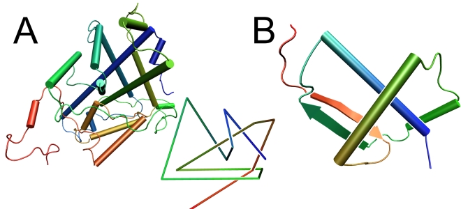
Figure 1. Protein crystal structure.
a: Crystal structure of α-haloacid dehalogenase DehI (PDB code 3bjx). The chain is composed of two homologous regions that form a pseudodimer and are connected by a proline-rich arc. The insert shows a reduced schematic representation of the protein. b: Crystal structure of the smallest knot discovered in an uncharacterized protein (PDB code 2efv). Both pictures were prepared with VMD [38].

