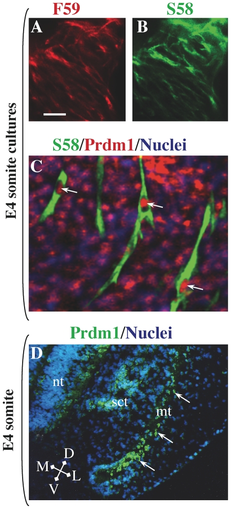
Figure 2. Expression of Prdm1 and fast and slow MyHC isoforms in E4 somite explant cultures and the somitic myotome.
Double immunofluorescence analysis for fast MyHC(s) with mAb F59 (A, red fluorescence) and slow MyHC(s) with mAb S58 (B, green fluorescence) showed that fast and slow MyHCs were co-expressed by all differentiated myocytes in somite cultures. In addition, Prdm1 immunostaining (Ab from Cell Signaling Technology) was found in the nuclei of all myocytes, as well as in many MyHC-negative cells (C, merged image, green fluorescence = S58, red fluorescence = Prdm1, blue fluorescence = nuclei). D. Prdm1 (green fluorescence, Ab from Abcam) was expressed throughout the myotome (mt, arrows) of a mature somite at the forelimb bud level at E4. Additional Prdm1 staining was found in some cells of the sclerotome (sct) and neural tube (nt). Arrows indicate Prdm1-positive nuclei within myocytes. Bar in Panel A = 40 µm for panels A and B); 15 µm for panel C; and 50 µm for panel D.

