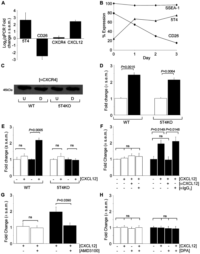
Figure 1. Differentiating mES cells show 5T4 dependent CXCL12 chemotaxis.
(A), Triplicate quantitative RT-PCR of WT-ES cells after 3 days differentiation with significant changes in 5T4, CD26 and CXCL12 mRNA but not CXCR4 respectively P = 0.014, 0.057, 0.81, 0.012 by Student's t-test. (B), Flow cytometry analysis of WT-ES cell differentiation 5T4 (triangles), CD26 (circles) and SSEA-1 (diamonds) (n>3 a single representative time course shown). (C), Western blot analysis of PAGE separated reduced WT or 5T4KO-ES cells either undifferentiated (U) or differentiating (D) probed with CXCR4 antibody. (D), Murine CXCL12 specific ELISA of conditioned medium from undifferentiated (white columns) and differentiating (black columns) WT and 5T4KO-ES cells. (E), Undifferentiated WT and 5T4KO-ES cells (white columns) exhibit no CXCL12 chemotaxis. Differentiating (black columns) WT, but not 5T4KO-ES cells, acquire significant chemotaxis. (F), CXCL12 chemotaxis in differentiating WT-ES cells (black columns) is blocked by an antibody against CXCL12; undifferentiated ES cells (white columns) show no chemotaxis. (G), Chemotaxis of differentiating WT-ES cells, (black columns) is blocked by a 2hr pre-incubation with 10 µM AMD3100; with no effect on undifferentiated WT-ES cells (white columns). (H), Undifferentiated (white columns) or differentiating (black columns) 5T4KO-ES cells show no change in chemotactic response in the presence of the CD26 inhibitor diprotin A (DPA, 10 µM).

