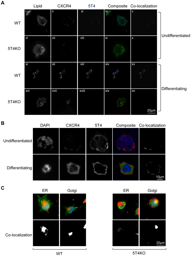
Figure 3. Cellular location of CXCR4, 5T4 in undifferentiated and differentiating WT and 5T4KO-ES cells.
(A), Shows lipid rafts in the membrane of all cells (i, vi, xi, xvi); CXCR4 is intracellular in undifferentiated WT-ES and all 5T4KO-ES cells (ii, vii) and cell surface 5T4 is only expressed on differentiation of WT-ES cells (xiv). The composite images (lipid = green, CXCR4 = red and 5T4 = blue) show co-localization of 5T4 and CXCR4 (purple) including in lipid rafts (white) in differentiating WT-ES cells (xiv) but no other cells (iv, ix, xix) (co-localized areas are shown in separate channel (v, x, xv, xx)). (B), RAd-m5T4 infection of 5T4KO-ES cells leads to cell surface expression of both 5T4 and CXCR4 only in differentiating cells but not in undifferentiated cells which are seen to co-localize (CXCR4 = green, 5T4 = red) in the composite (yellow)(co-localized areas are shown in separate channel). RAd-GFP showed no effect on CXCR4 expression (not shown). (C), Upper panels, Double labeling of WT or 5T4KO-ES cells with either NBD C6-Ceramide (Golgi) or Endotracker (ER) (both red) shows that in the absence of 5T4, CXCR4 (green) accumulates predominately in the Golgi and to a lesser extent the smooth ER (yellow) whereas cell surface labeling is apparent only in the differentiating WT-ES cells)(lower panels: co-localized areas are shown in separate channel).

