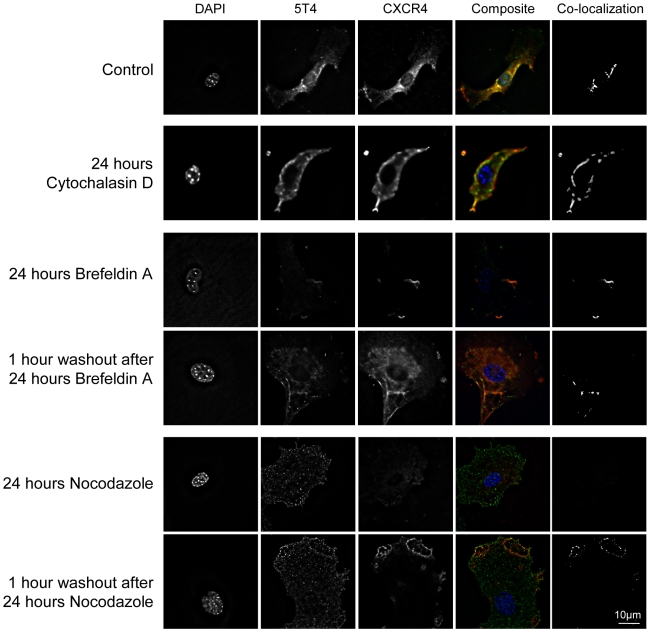
Figure 7. Effects of cytoskeleton, microtubule and Golgi disruption on the co-localisation pattern of 5T4 and CXCR4.
Primary murine embryonic fibroblasts were assessed for their pattern of 5T4 and CXCR4 expression by immunofluorescence following 24 hours disruption of either the cytoskeleton (cytochalasin D), Golgi (brefeldin A) or microtubules (nocodazole) and 1 hour after drug washout. Cell surface expression of 5T4 (green) and CXCR4 (red) with regions of co-localization of the two antigens (seen as yellow) (also shown by co-localization analysis) are depicted.

