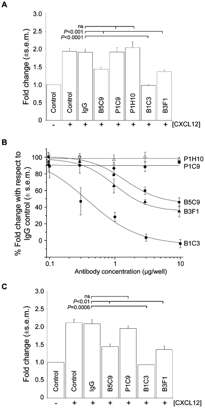
Figure 9. Inhibition of chemotaxis by monoclonal antibodies recognizing 5T4.
(A), The chemotactic migration exhibited by differentiating WT-ES cells towards CXCL12 was abolished in the presence of the m5T4 specific mAb B1C3 (10 µg) but not in presence of mAb P1C9 or P1H10 (10 µg) or an irrelevant control antibody (10 µg). MAbs B3F1 and B5C9 (10 µg) reduced the chemotactic response. (− = no CXCL12, + = 10ng CXCL12). (B), MAb dose response of inhibition of chemotaxis towards CXCL12 in differentiating WT-ES cells. (C), The chemotactic migration exhibited by primary WT MEF was abolished in the presence of the m5T4 specific mAb B1C3 (10 µg) but not in presence of mAb P1C9 (10 µg) or an irrelevant control antibody (10 µg). MAbs B3F1 and B5C9 (10 µg) reduced the chemotactic response.

