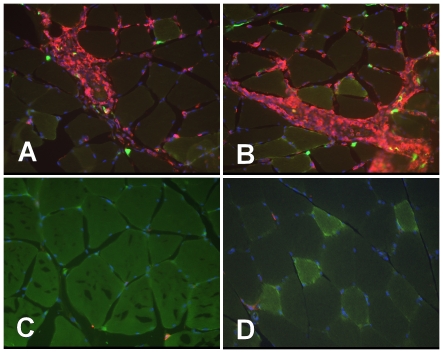
Figure 5. Histochemistry of rat muscle injected with GFP lentiviral vector.
Wistar rats were injected with a lentiviral GFP expression vector (A-C) or saline control (D). (A,B) Areas with remnants of GFP expressing cells (green) show co-localization with CD45 stained (red) infiltrating immune cells. (C) Areas without GFP expression within the same piece of tissue are devoid of infiltrating immune cells. (D) Control injected muscle is negative for both GFP expression and infiltrating immune cells.

