
Fig. 1.
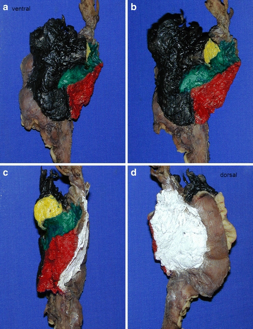
a–d Stained surfaces of pancreatic head resection specimen (black—ventral; white—dorsal; yellow—pancreatic RM; green—groove of superior mesenteric vein; red—mesopancreatic RM)
Official websites use .gov
A
.gov website belongs to an official
government organization in the United States.
Secure .gov websites use HTTPS
A lock (
) or https:// means you've safely
connected to the .gov website. Share sensitive
information only on official, secure websites.
a–d Stained surfaces of pancreatic head resection specimen (black—ventral; white—dorsal; yellow—pancreatic RM; green—groove of superior mesenteric vein; red—mesopancreatic RM)