Figure 6.
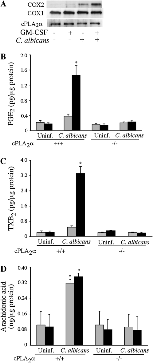
GM-CSF priming enhances COX2 expression and prostanoid production in response to C. albicans. (A) Levels of COX2, COX1, and cPLA2α were determined by Western blot analysis of cell lysates prepared from unprimed (shaded bars) and GM-CSF–primed (solid bars) cPLA2α+/+ alveolar macrophages stimulated with or without C. albicans (moi, 2) for 6 hours. The Western blot results are representative of three independent experiments. (B–D) Unprimed and GM-CSF–primed cPLA2α+/+ and cPLA2α−/− alveolar macrophages (Balb/c) were infected with C. albicans (moi, 2) for 6 hours. Culture medium was collected and analyzed for levels of (B) PGE2, (C) TXB2, and (D) AA by mass spectrometry. Data are the average ± SEM of three experiments. GM-CSF–primed cPLA2α+/+ macrophages release significantly more (*P < 0.05) PGE2 (B) and TXB2 (C) than unprimed cPLA2α+/+ macrophages, and significantly more than cPLA2α−/− macrophages in response to C. albicans infection. C. albicans–infected cPLA2α+/+ macrophages (primed and unprimed) release significantly more (*P < 0.05) AA than cPLA2α−/− cells.

