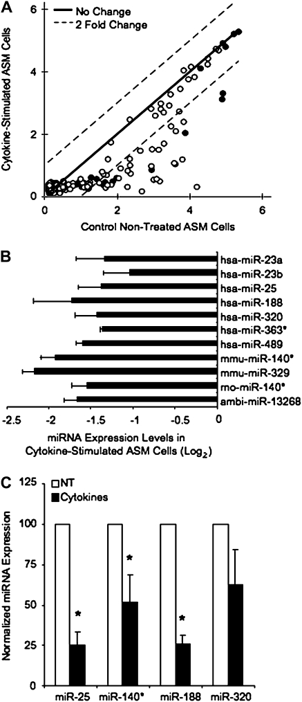
Figure 2.
Cytokine stimulation affects miRNA expression in human ASM cells. Human ASM cultures were grown to confluence and serum-starved for 48 hours before cytokine treatment with 10 ng/ml IL-1β, TNF-α, and IFN-γ for 24 hours. RNA was isolated and miRNA arrays hybridized and analyzed as described. (A) A scatter plot summarizes the mean signal intensity on a log2 scale from the 134 miRNA expressed in two different ASM cultures (shown in solid and open circles) assayed in duplicate. Dotted lines indicate a 2-fold expression change between cytokine-stimulated and nontreated cultures; the solid line indicates no changes. (B) Ratios of miRNA expression in cytokine-stimulated versus nontreated cultures were generated from miRNA array analysis. Normalized ratios of the 11 miRNA down-regulated ≥ 2-fold with cytokine treatment are shown. (C) TaqMan miRNA expression assays were used to verify changes in miRNA expression seen on the arrays. Human ASM cultures were treated as above, RNA extracted with TRIzol, and miRNA further purified for cDNA synthesis. RT-PCR were performed with TaqMan miRNA assays using 250 ng RNA enriched for miRNA. miRNA expression was normalized to U6 expression in each sample. Data is expressed as the % change in miRNA expression from nontreated cultures (100%) (n = 3–6 ± SEM). *Statistically significant difference from control, nontreated cultures (P < 0.05).

