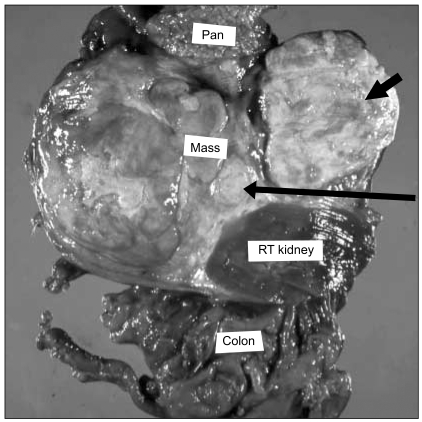
Fig. 3.
On sectioning, the tumor mass shows a poorly circumscribed solid cut surface with multifocal necrosis, calcification and discrete intratumoral nodules of varying sizes. The right kidney is partially encased and the mass has grossly infiltrated the surrounding duodenum (thick arrow), the inferior vena cava wall (thin arrow), the pancreas and the colon.

