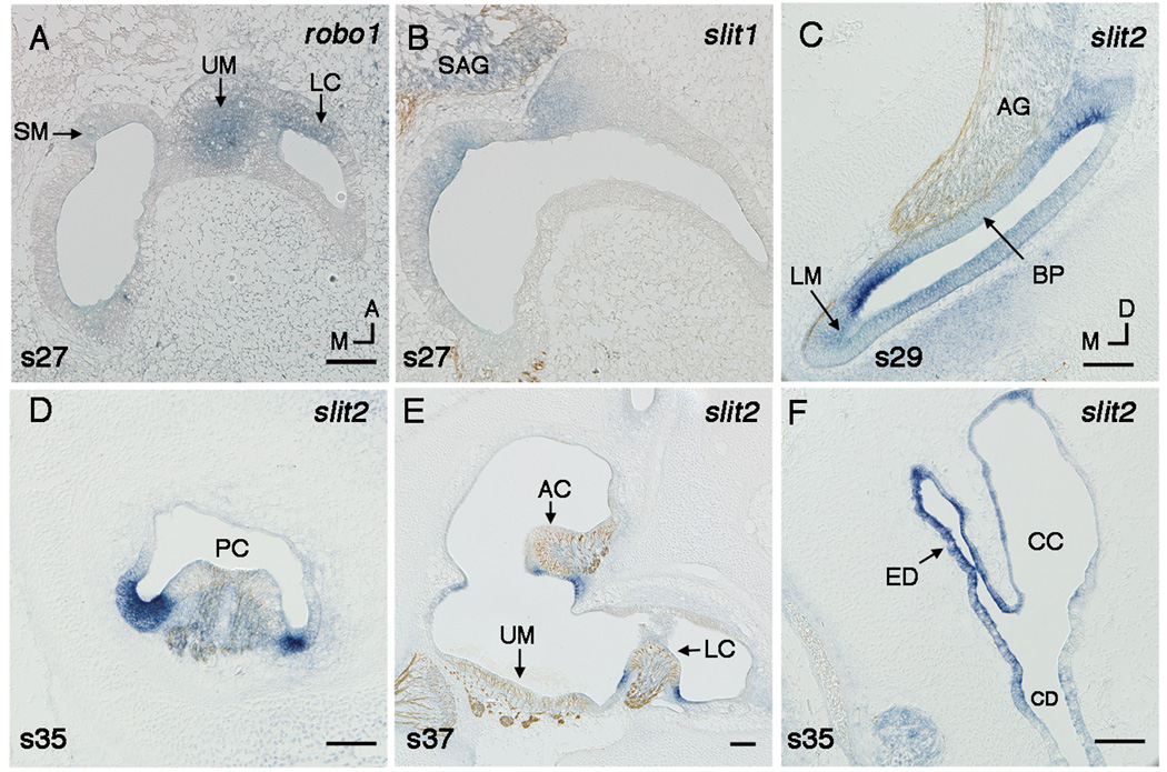
Fig. 3.
A,B: Robo1 (A) and slit1 (B) in the utricular macula at stage 27 shown by in situ hybridzation on horizonal sections. C–E: Slit2 flanks sensory territories shown in transverse sections. Sections (B,C,E) are double labeled with a neurofilament antibody. Slit2 flanks the auditory basilar papilla (BP) at stage 29 (C). Slit2 flanks the vestibular sensory epithelia, including the posterior crista (PC) at stage 35 (D), and the anterior crista (AC), and the lateral crista (LC) at stage 37 (E). Slit2 is expressed in the endolymphatic duct (ED) at stage 35 (F). Scale Bars, 100µm. Orientation: A, anterior; D, dorsal; M, medial. Abbreviations: CC, common crus; CD, cochlear duct; SM, saccular macula; SAG, statoacoustic ganglion; UM, utricular macula.

