
Figure 1.
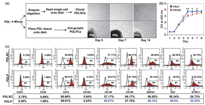
Isolation of human PDLPs. (a) Comparison procedure of isolating PDLPs and PDLSCs from periodontal tissues and morphology of culture expanded cells at 5, 7, and 14 days (magnification 100×). (b) At days 1–3, PDLPs showed similar proliferation rate as PDLSCs, but showed an elevated proliferation from day 4 to 7. After day 8, PDLPs have similar proliferation rate as PDLSCs in the culture. (c) Cytometric flow analysis indicated that PDLPs have similar surface molecule phenotype as PDLSCs derived from same donor including negative for CD34 and CD45, and positive for CD73, CD105, CD146, CD166, SSEA4, 3G5, and STRO-1 (*P < 0.05; **P < 0.01). Experiments were repeated three times