
Figure 4.
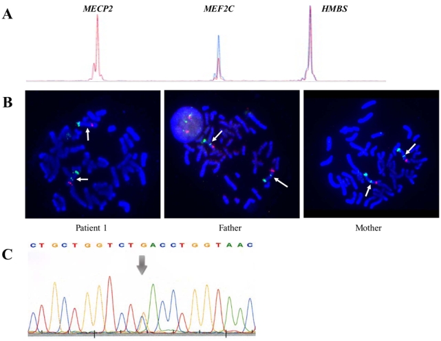
Confirmation of the 5q14 deletion by QMPSF and FISH in case 1. (A) Electropherogram in case 1 (in red) was superimposed to that of a normal female (in blue) by adjusting to the same level the peaks obtained for the HMBS and MECP2 control amplicons. Case 1 is a female. The Y axis displays fluorescence in arbitrary units, and the X axis indicates the size in bp. Heterozygous 5q14 deletions are easily detected by 50% reduction of the MEF2C peak in the patient compared to a normal control. (B) Results of two-colour FISH analysis in case 1 with RP11-1006G2 labelled with SpectrumGreen in combination with Cridu-chat syndrome probe used as control (5p15.2, Spectrum Green, 5q31 EGR1 Spectrum Orange, Abbott). Note that RP11-1006G2 is lacking on one chromosome 5 in the patient and is present on both homologous chromosomes in the parents, demonstrating de novo origin of the deletion. (C) Sequencing DNA chromatogram in case 7. Arrow indicates the c.683 C>G mutation within exon 7 of MEF2C.