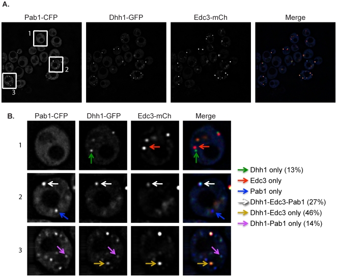
Figure 2. Dhh1 localizes to both P-bodies and stress granules following glucose deprivation stress.
(A) A strain harboring a Dhh1-GFP integration was transformed with a plasmid containing Edc3-mCherry and Pab1-CFP and assayed for GFP co-localization with mCherry and/or CFP upon glucose deprivation. (B) A closer view of three sample cells, labeled accordingly in A, is shown. Dhh1 co-localized with Edc3-mCherry (gold arrows), Pab1-CFP (purple arrows), and both Edc3-mCherry and Pab1-CFP (white arrows). Dhh1 was also found in independent foci (green arrows).

