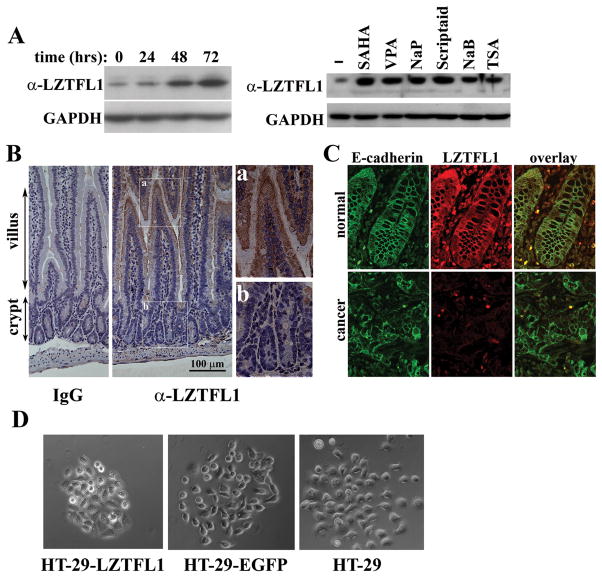
Figure 5. LZTFL1 expression is up-regulated upon cell differentiation.
(A) Left panel, HT-29 cells were cultured in the presence of NaB for the indicated times. The LZTFL1 expression level was measured by Western blot. Right panel, HT-29 cells were cultured for three days in the absence (−) and presence of the HDAC inhibitors, suberoylanilide (SAHA), Valproic acid (VPA), sodium propionate (NaP), scriptaid, NaB, and Trichostatin A (TSA). Cell lysates were subjected to Western blot analysis with anti-LZTFL1 antibody. (B) Immunohistochemical staining of IgG (negative control) and LZTFL1 in the mouse small intestine. There is a graded LZTFL1 staining along the crypt-villus axis (middle panel, 20×). Inset a shows a robust LZTFL1 staining in the apex of villus (40×). Inset b shows absence of LZTFL1 staining in the crypt. (C) Confocal microscopy images of human normal colonic tissue (upper panels) and colorectal carcinoma (lower panels) stained with E-cadherin (green) and LZTFL1 (red). The expression of LZTFL1 is overlapping with that of E-cadherin at the plasma membrane in normal colonic epithelial cells. (D) Parental and pires-EGFP and pLZTFL1-ires-EGFP transfected HT-29 cells were seeded with a low density to allow them to grow as discrete colonies. PMA (0.5 μg/ml) was added to the cell to induce cell scattering. Micrographs were taken 8 hrs after addition of PMA.

