
Figure 3.
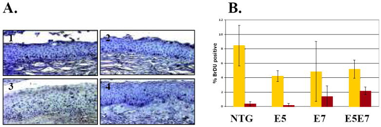
Characterization of the proliferative index in cervical epithelia. A. Representative pictures of BrdU-specific immunohistochemical staining of cervical epithelium from nontransgenic (1), E5 (2), E7 (3), and E5E7 (4) mice. B. Quantification of BrdU-labeling index of cells within distinct layers of cervical epithelia. The average percentage of basal (yellow) and suprabasal (red) BrdU-positive cells was obtained from ten (× 40) microscope fields per mouse. An average of at least three mice per genotype were used to calculate the percentage. The difference in % suprabasal DNA synthesis between E5E7 and E7 cervical epithelium was statistically significant (p=0.02, two-sided Wilcoxon rank sum test).