
Figure 4.
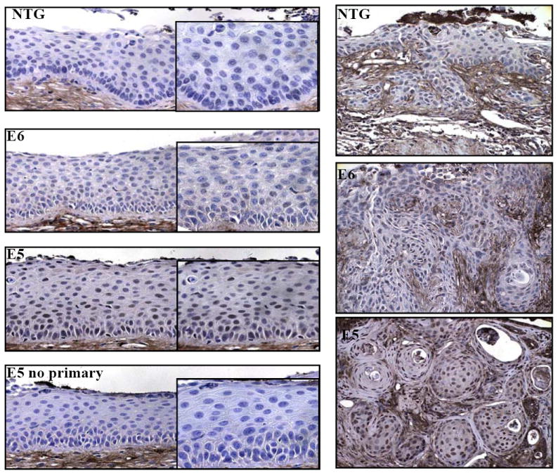
ERK1/2 activation in transgenic mice treated 9 months with exogenous estrogen. Sections from nontransgenic, E6, and E5 transgenic mice were immunohistochemically stained for the presence of phosphorylated ERK1/2. Shown are representative pictures of cervical epithelium (left panels) and tumors (right panels) from nontransgenic, E6, and E5 transgenic mice treated with exogenous estrogen for 9 months. Right panels insets: higher magnifications of epithelium. Bottom right panel: no primary antibody control.