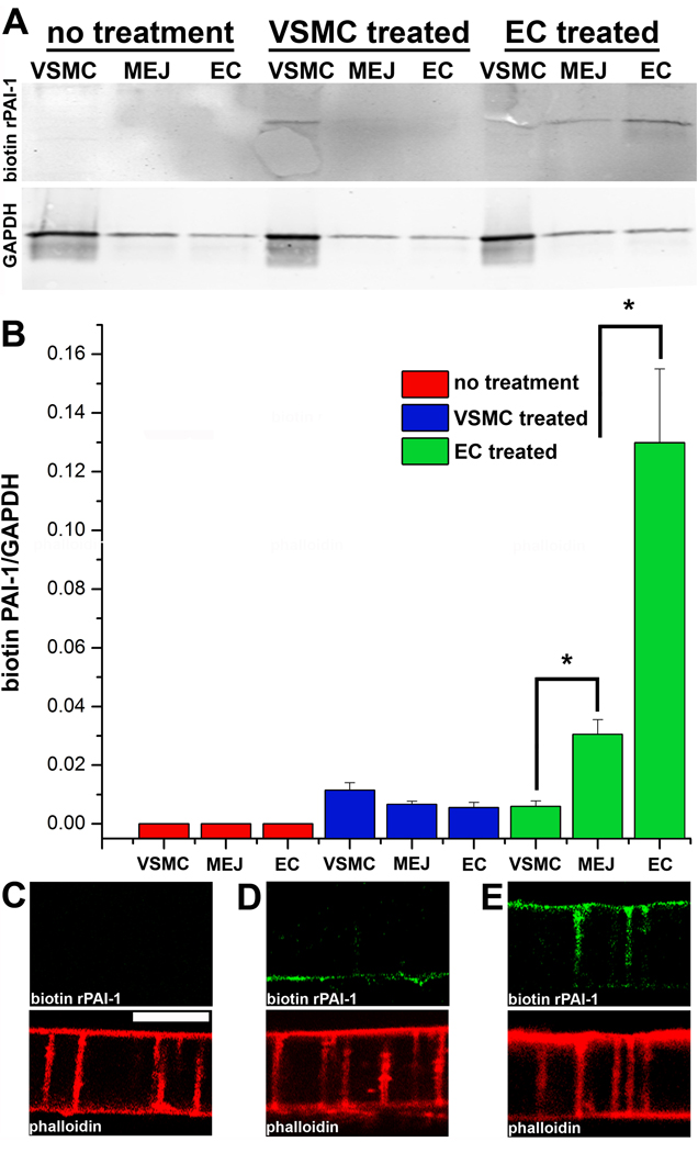
Figure 6. Uptake and expression of biotin-conjugated rPAI-1 at the MEJ.
Immunoblots of VSMC, MEJ and EC fractions isolated from the VCCC stained with streptavidin. Three experimental paradigms were tested: no application of biotin-conjugated rPAI-1, application of biotin-conjugated rPAI-1 (0.1µg) to the VSMC monolayer and application of biotin-conjugated rPAI-1 (0.1µg) to only the EC monolayer. GAPDH is shown directly below each condition (A). Normalized quantification of protein expression for biotin-conjugated rPAI-1 for each fraction and experimental paradigm is given in (B). Immunocytochemistry on transverse sections of the VCCC for biotin-conjugated rPAI-1 using streptavidin (green) and F-actin (with phalloidin, red) for the following experimental paradigms: no application of biotin-conjugated rPAI-1, application of biotin-conjugated rPAI-1 (0.1µg) to the VSMC monolayer and application of biotin-conjugated rPAI-1 (0.1µg) to only the EC monolayer). For all images, VCCC were treated 30 minutes prior to isolation. Bar in C is 10 µm and representative for images (C-D). *p<0.05. For A–B, n=4.

