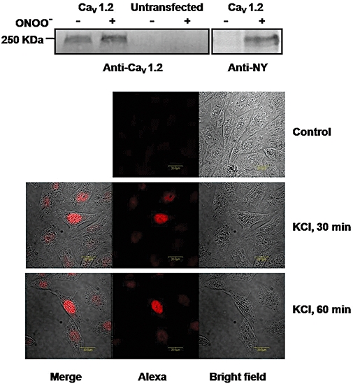
Figure 2.

Protein tyrosine nitration of the Ca2+ channel following in vitro treatment with peroxynitrite (ONOO-) in hCav1.2b-transfected CHO cells. (A) Immunoblot with anti-Cav1.2 antibody (left) and anti-nitrotyrosine antibody (right). Protein samples were immunoprecipitated with the Ca2+ channel antibody before immunoblot. Treatment with peroxynitrite enhanced tyrosine nitration of hCav1.2b. (B) Expression of phospho-CREB in hCav1.2b-transfected CHO cells detected by anti-pCREB antibody (1:50 dilution). Staining in the nucleus was observed in hCav1.2b transfected cells treated with 80 mM KCl for either 30 min or 60 min but not in the absence of depolarization (top panel). An Alexa Fluor 568 conjugated antibody (1:1000 dilutions) was used as the secondary antibody. Representative images from three experiments are shown. CREB, cyclic AMP response element binding protein.
