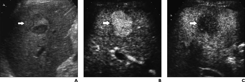
Fig. 1.
53-year-old man with liver cirrhosis related to hepatitis C virus infection and with poorly differentiated hepatocellular carcinoma.
A, Unenhanced longitudinal sonogram shows heterogeneous nodule (arrow) with diameter of 2.5 cm.
B and C, Contrast-enhanced Cadence Contrast Pulse Sequencing (CPS; Acuson Sequoia, Siemens Medical Solutions) longitudinal sonograms show nodule (arrow) is homogeneously hypervascular 25 seconds after microbubble injection during arterial phase (B) and hypovascular in comparison with adjacent liver parenchyma during portal venous phase (C).

