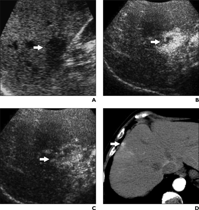
Fig. 6.
41-year-old woman with liver cirrhosis and intrahepatic arterioportal shunt.
A, Unenhanced oblique sonogram shows one nodule (arrow) with diameter of 1.5 cm.
B and C, Contrast-enhanced Coherent Contrast Imaging (CCI; Acuson Sequoia, Siemens Medical Solutions) oblique sonograms after microbubble show injection nodule (arrow) is encompassed by diffuse hypervascular appearance persisting during arterial (B) and portal venous (C) phases.
D, Contrast-enhanced CT scan of nodule (arrow) shows clear hypervascularity in arterial phase. Because it appeared solid on unenhanced sonography, nodule was biopsied and later surgically resected.

