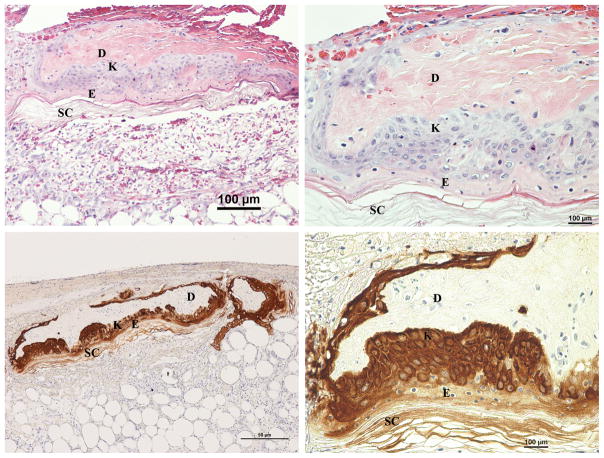
Fig. 4.
Day 4 H&E (Above left & right) and Cytokeratin (Below left & right) stained sections of wounds with inverted meshed skin graft in the wet environment. H&E sections show an inverted section of the graft with proliferation of the basal keratinocytes throughout its entire length. The upper layers of the original epidermis are not viable at this point. Migrating keratinocytes can be seen moving upward toward the wound surface. D, original dermis, E, original epidermis and SC, original stratum corneum of the skin graft; K, proliferating keratinocytes.

