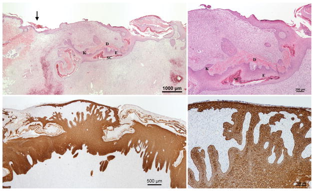
Fig. 5.
Day 9 H&E (Above left & right) and Cytokeratin (Below left & right) stained sections of wounds with inverted meshed skin graft in the wet environment. (Above left & right). Proliferation of keratinocytes arising from the skin graft and moving upward to form the new epidermis can be seen. There is not yet connection between the migrating epithelium originating from the surrounding unwounded skin and the migrating keratinocytes from the skin graft. The arrow indicates the wound margin. The dermis and epidermis of the skin graft appear completely separated. (Below left) The newly formed epithelium at this stage is thick. Some segments of the skin graft are being eliminated from the surface. (Below right) An advancing layer of migrating keratinocytes on the surface with several connecting bridges of keratinocytes from the deeper portions of the wound.
D, original dermis, E, original epidermis and SC, original stratum corneum of the skin graft; K, proliferating keratinocytes.

