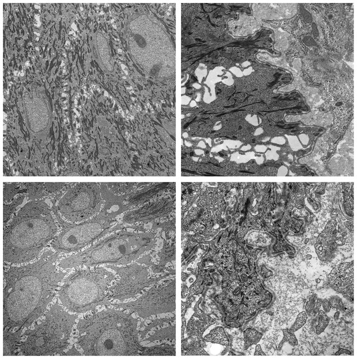
Fig. 9.
Electron microscopy of the wounds treated in the wet environment on day 9. Wounds treated with regular meshed graft show well developed basement membrane (Above right) and hemidesmosomes (Above left). Wounds with inverted meshed graft also revealed well-developed basement membrane (Below right) but much fewer hemidesmosomes (Below left).

