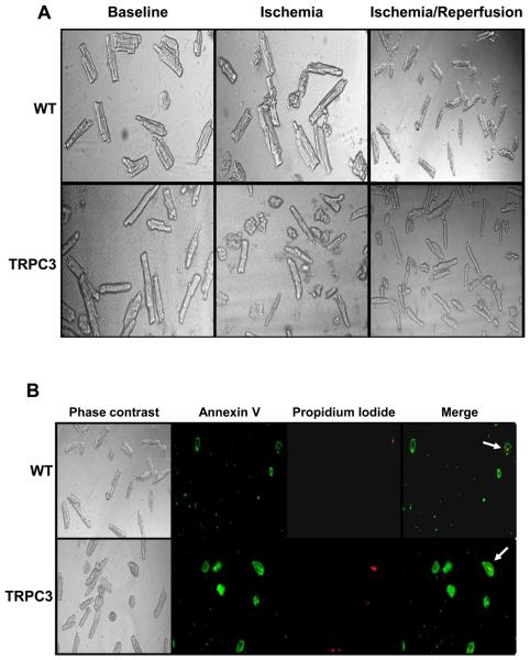
Figure 2.
A) Bright field phase contrast images of cardiomyocytes from wild-type (WT) and TRPC3 transgenic mice at baseline, at the end of 90 min ischemia and after 90 min ischemia and 3 hours reperfusion; B) cardiomyocytes from wild-type (WT) and TRPC3 transgenic mice after 90 min ischemia and 3 hours reperfusion showing Annexin V and propidium iodide staining. Arrows in merged image indicate necrotic cells, staining positive for both Annexin V and propidium iodide.

