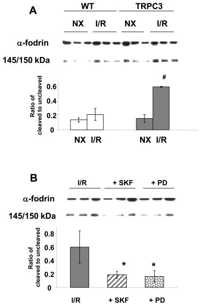
Figure 5.
A) Total and cleaved α-fodrin (145/150kD) in cardiomyocytes from wild-type (WT) and TRPC3 transgenic mice under normoxic conditions (NX) and following 90 min ischemia and 3 hours reperfusion (I/R); B) total and cleaved α-fodrin (145/150kD) following 90 min ischemia and 3 hours reperfusion (I/R) in untreated cardiomyocytes from TRPC3 transgenic mice and in TRPC3 cardiomyocytes treated with the CCE inhibitor SKF96365 (SKF, 0.5 μM) and the calpain inhibitor PD150606 (PD, 25 μM). Upper panels are representative immunoblots and the lower panels are mean densitometric data from 3 individual experiments. # = p < 0.05 vs. TRPC NX and WT I/R; * = p < 0.05 vs. I/R.

