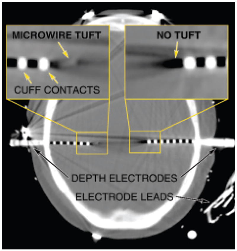
Fig. 4.

Computed tomography scan showing the implanted depth electrodes. Depth electrodes similar to ones that would be appropriate for use in an LGN-based visual prosthesis are already in clinical use during preparation for surgical treatment of epilepsy such as shown in this image obtained in a patient with bilaterally implanted hippocampus. Left inset: A depth electrode that combines traditional cuff-style contacts with a central bundle of microwires exiting distally. Right inset: A traditional depth electrode without the central bundle.
