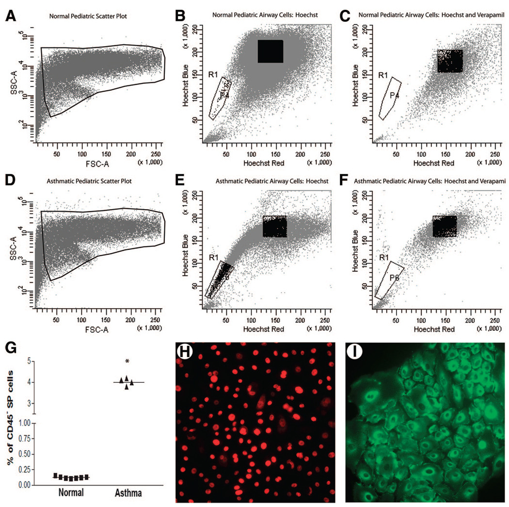
Figure 5.
Identification of lung SPs in asthmatic airways. Representative fluorescence-activated cell sorting FSC and SSC plots of airway epithelial SP cells in normal and asthmatic pediatric patients ([A, D], region R1) demonstrate a distinct SP that represented 4.01% of the epithelial population in asthmatic pediatric airways ([B], region R1) and 0.11% in normal pediatric airways ([E], region R1). Detection of SP cells was inhibited in the presence of 50 µg/ml verapamil ([C, F], region R1). (G): Mean percentages of SP cells for all normal (■) and asthmatic (▲) individuals in the study; * indicates p < .05. (H, I): Representative images demonstrating ΔNp63 and E-cadherin staining in asthmatic CD45− SP colonies. Abbreviations: FSC, forward scatter; SP, side population; SSC, side scatter.

