
Figure 4.
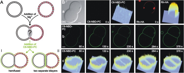
Lipids in the outer leaflet cannot enter the HD. C6-NBD-PC was added to pairs of GUVs with sequestered Rh-HA peptides. After insertion of the lipid analog in the outer leaflet, labeling of the contact region was studied by following the lateral distribution of the NBD fluorescence. (A) Sketch of C6-NBD-PC localization. In case of HD formation no redistribution of the lipid analog to the HD is observed (I) whereas the adhesion region becomes labeled when it is formed by two separated bilayers (II). (B) Lateral distribution of C6-NBD-PC observed by confocal fluorescence microscopy. (a) Images of a GUV pair before addition of C6-NBD-PC (t = 0). From left to right: Differential interference contrast; distribution of C6-NBD-PC (green); intensity profile of NBD fluorescence; distribution of Rh-labeled peptide (red); intensity profile of rhodamine fluorescence; (b) distribution of C6-NBD-PC and (c) corresponding intensity profile at various times after addition of C6-NBD-PC. Scale bar = 5 μm.