
Figure 1.
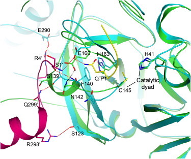
Active center of the SARS-CoV Mpro. The interactions between the P1 substrate-binding subsite from chain A (cyan) and the N-finger and domain III from chain B (magenta) of SARS-CoV Mpro (PDB code 1UK4) are shown. The substrate analog and the side chain of the Gln-P1 residue are yellow. The R298A monomeric structure (green) (PDB code 2QCY) is superimposed on the wild-type chain A. The dashed lines indicate hydrogen bonds for 1UK4 (red) and 2QCY (black). This figure was produced using PyMOL (35).