
Figure 7.
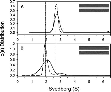
Sedimentation velocity experimental values for Glu-166 mutants E166A (A) and E166A/R298A (B) in D2O. The protein concentration was 0.33 mg/ml. The distributions in D2O are represented by solid lines and those in D2O with 600 μM TQ6-pNA substrate by dashed lines. The left vertical dotted line indicates the monomer position and the right, the dimer position. (Insets) Residual bitmaps for mutants without (upper) and with (lower) substrate.