Figure 3.
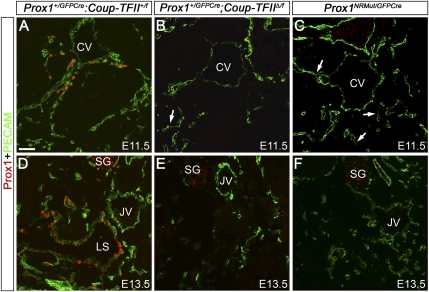
Interaction between Coup-TFII and Prox1 is required to maintain Prox1 expression in LECs. (A) At E11.5, Prox1-expressing LECs (red) are seen in and around the anterior cardinal vein (CV) of Prox1+/GFPCre;Coup-TFII+/f embryos. (B) Just a few Prox1+ LECs (arrow) are seen in Prox1+/GFPCre;Coup-TFIIΔ/f littermates. (C) E11.5 Prox1NRMut/GFPCre embryos expressing a form of Prox1 mutated in the nuclear hormone receptor-binding site also have a reduced number of LECs (arrows). (D) At E13.5, the lymph sacs (LS) lined by Prox1+ LECs are seen in control embryos. However, at this stage, no LECs are seen in Prox1+/GFPCre;Coup-TFIIΔ/f (E) or Prox1NRMut/GFPCre (F) embryos. PECAM is shown in green. The neural tube is oriented toward the left side of all panels. (JV) Jugular vein; (SG) sympathetic ganglia. Bar, 50 μm.

