Fig. 4.
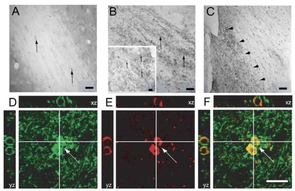
Nogo-A expression in ipsilateral white matter tracts in sham- and brain-injured animals. (A) In the external capsule (EC) of sham-injured animals, immunoreactivity for Nogo-A was observed in oligodendrocytes along the white matter fiber tracts (arrows, scale bar = 100 μm). (B) FP brain injury induced a marked increased expression of Nogo-A in the EC ipsilateral to the injury at all time-points evaluated (arrows, scale bar = 100 μm). Insert shows higher magnification of the EC of another brain-injured animal, showing an increased number of Nogo-A (+) cells (arrows, scale bar = 50 μm). (C) Example of increased immunostaining for Nogo-A at 7 days post-injury in the medial, but not lateral part (arrowheads) of the fimbria ipsilateral to the injury, scale bar = 200 μm. (D–F) Confocal reconstruction of the EC of a FP brain-injured animal. An increased number of RIP (+) (D, green)/Nogo-A (+) (E, red)-colabeling cells (F; merged cells shown in yellow, arrows) were observed. Scale bar = 50 μm.

