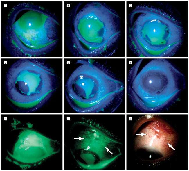
Figure 3.
Unique healing pattern under ProKera (Bio-Tissue, Inc, Miami, Florida). Case 4, grade II, with superior limbal ischemia, total corneal and limbal epithelial defects, and associated perilimbal conjunctival epithelial defect (A and G), showed rapid perilimbal conjunctival healing and epithelialization of the cornea from the 4-o’clock position 5 days after insertion of ProKera (B). At day 7, the expanded epithelial mass at the 4-o’clock position had circumferentially moved to the limbal region while a limbal epithelial mass emerged from the 7-o’clock position where the conjunctival defect had closed (C). At day 10, the epithelial mass from the 7-o’clock position had enlarged (D), and again moved circumferentially toward the limbal region at day 12 (E). At day 17, the corneal surface was completely healed (F). A large conjunctival epithelial defect that extended to the superior bulbar area was noted before amniotic membrane patching (G). This defect was not healed by day 14 in the area outside of ProKera (H, arrows mark the skirt), and by day 17 had evolved into a granulation tissue where hyperemic blood vessels emanated from the fornix but not from the amniotic membrane–covered limbus (I).

