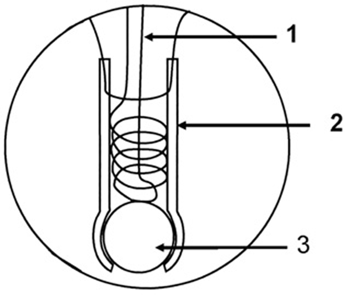
Fig. 1.
Diagram of the membrane inlet which is inserted in an anaerobic cell for mass spectrometric measurements: (1) Wire helix for support of the Silastic tubing; (2) Silastic tubing approximately 1 cm in length; (3) glass bead to seal the Silastic tubing. Taken from Ref. [9].

